Eye exams are professional screenings used to evaluate the health of the eye and diagnose vision impairments and disease.
Each is comprised of a series of several tests that analyze various functions of the eye, such as color differentiation, distance vision, and peripheral awareness. A comprehensive eye exam can identify vision complications during their earliest stages, providing eye doctors the opportunity to treat them more easily. It should be part of your general wellness checkups including annual physicals, teeth cleanings and dental visits, and eye exams.
A comprehensive eye exam will consist of:
- Visual acuity testing to measure the clarity of your sight
- Refractive evaluation to diagnose refractive errors
- Visual field testing to measure your peripheral vision
- Observing your pupils’ response to light to detect neurological disorders
- Color vision analysis to identify an inability to distinguish certain colors
- Examination of the back of your eye, retina, and underlying blood vessels
- Glaucoma testing to measure intraocular fluid pressure
- Eye muscle testing to identify weak eye movements
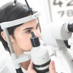
- Corneal exam to evaluate the health of the cornea, iris, lens and surrounding tissues
Did you know?A periodic eye exam can reveal underlying health issues long before other symptoms are present. In addition to identifying vision complications and eye disease, a comprehensive eye examination can also reveal conditions like high blood pressure, diabetes, a stroke, or even a brain tumor. The American Optometric Association recommends that adults under age 60 undergo a comprehensive eye exam at least once every one to two years. At-risk patients and patients over age 60 should get an eye exam annually. |
TECHNOLOGY
Optical Coherene Tomography 3D
The Topcon 3D OCT-1 Maestro System provides the latest diagnostic technology available to allow the doctors to examine different layers of the retina.
This instrument looks microscopically at the layers of the retina for early signs of macular degeneration and diabetic macular edema.
It is on the cutting edge of technological advancements for viewing the retina and it allows our doctors to see underneath the retina and identify problems that cannot be seen with the naked eye. This is also effective for early detection of glaucoma.
![]()
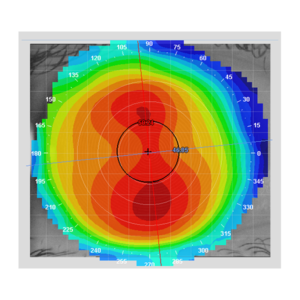
TOPOGRAPHER
New Optix Optometry is equipped with the latest corneal topographer which is a non-invasive medical imaging technique for mapping the surface curvature of the cornea. This helps the doctor diagnosis and treat a number of corneal conditions including keratoconus and high astigmatism by fitting speciality lenses. The topographer is also a valuable instrument used to properly fit Ortho K lenses.
WIDE FIELD RETINAL CAMERA
Our retinal camera takes a quick and painless digital image of our retina, the tissue responsible for allowing you to see. This technology assists in determining the health of your eyes and can help identify and manage diseases like diabetes, glaucoma, macular degeneration, and many more conditions that may affect the health of your eyes. All our locations are equipped with a wide field retinal camera.

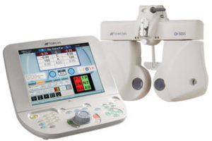
DIGITAL AUTO PHOROPTER
The days of ” Which is clearer, 1 or 2″ are long gone. You can now compare the clarity side by side. This extremely accurate technology makes the process of finding your prescription faster and more enjoyable.